Insights into the role of PAD2-mediated histone citrullination in pancreatic cancer progression
Knockdown of peptidyl-arginine deiminase 2 gene in pancreatic cancer cells leads to decreased cell proliferation, finds new study.
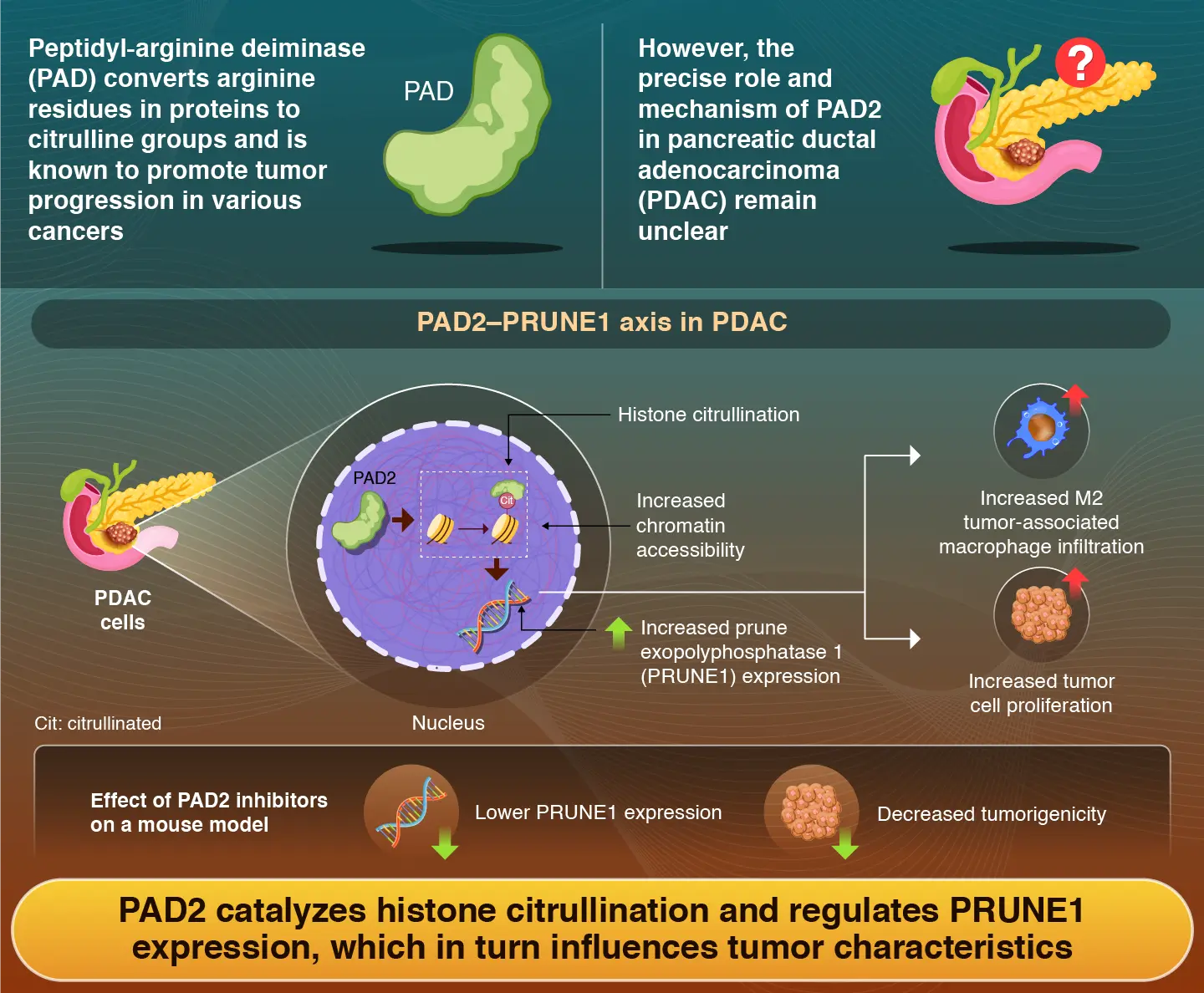
Peptidyl-arginine deiminase 2 (PAD2) enzyme converts arginine amino acid residues in histone proteins into citrulline groups and promotes tumor cell proliferation in pancreatic ductal adenocarcinoma, report researchers from Institute of Science Tokyo, Japan. Administration of PAD inhibitors reduced PRUNE1 expression and suppressed tumor cell proliferation in both pancreatic cancer cell lines and mouse models. The study thus lays the foundation for future anticancer therapies targeting PAD2 enzymatic activity.
Role of Peptidyl-Arginine Deiminase 2 in Pancreatic Ductal Adenocarcinoma Progression

Umemura et al. (2025) | Molecular Cancer Research | 10.1158/1541-7786.MCR-24-1095
Pancreatic ductal adenocarcinoma (PDAC), a type of pancreatic cancer, can be clinically challenging to treat and is often associated with poor patient outcomes. Cancer metastasis, or the ability to spread to other organs, and the development of resistance to anticancer therapy are well-known mechanisms that contribute to PDAC-related health complications. To limit the aggressive growth of cancer and develop novel therapeutics against PDAC, scientists around the globe have systematically investigated the genetic and protein expression profiles of tumor cells.
Recent reports indicate that the peptidyl-arginine deiminase (PAD) enzyme is widely expressed in many cancers and is capable of regulating gene expression in tumor cells. The PAD family of enzymes specifically converts arginine amino acid residues in histone proteins into citrulline groups—a process called histone citrullination. This chemical modification of histone proteins results in increased access to deoxyribonucleic acid strands, thereby aiding tumor cell proliferation. However, the precise role and molecular mechanism of PAD2 in PDAC are yet to be elucidated.
To address this research gap, a team of scientists led by Professor Shinji Tanaka from the Department of Molecular Oncology, Institute of Science Tokyo (Science Tokyo), Japan, along with Lecturer Yoshimitsu Akiyama and graduate student Kentaro Umemura, conducted a new study. They employed advanced genetic manipulation techniques in laboratory-grown cancer cells and mouse models to assess the mechanistic effects of PAD2 in PDAC. Their findings were made available online on June 12, 2025, and published in the journal Molecular Cancer Research on June 26, 2025.
During the initial stages of their study, the researchers conducted immunohistochemistry staining of tissue samples obtained from patients with PDAC to detect histone citrullination. Their analysis revealed that histone citrullination was more prevalent in PDAC compared to normal pancreatic tissue and was associated with poor overall survival.
Subsequently, the scientists shifted their attention to establishing the functional role of PAD2 in PDAC. They generated PAD2-overexpressing (OE) and PAD2-knockdown (KD) cell lines using genetic manipulation techniques. The results of cell proliferation assays showed that PAD2-KD cells lacking PAD2 had decreased cell proliferation, while PAD2-OE cells exhibited a significant increase in the cell proliferation rate.
To identify downstream target genes affected by PAD2 activity, the research team conducted RNA-sequencing analysis of PAD2-KD cells. They found that eight genes were significantly downregulated in PAD2-KD cells. Interestingly, among the eight genes, prune exopolyphosphatase 1 (PRUNE1) and E2F transcription factor 1 were upregulated in PAD2-OE cells, suggesting that PAD2 directly influences their gene expression.
Finally, to validate their findings, the scientists injected PAD2-OE cells or PAD2-KD cells into mice to generate tumors. In mice injected with PAD2-OE cells, the formation of tumors was much higher, and protein staining analysis revealed increased levels of histone citrullination. Notably, advanced immunofluorescence-based staining demonstrated that tumor-infiltrating “M2” macrophages were greatly increased within PAD2-OE tumors in mice.
“Treatment of PAD2-OE cells and other pancreatic cancer cell lines with Cl-amidine, a general PAD inhibitor, and AFM-30a, a PAD2-specific inhibitor, led to lower PRUNE1 expression. Moreover, intraperitoneal administration of Cl-amidine inhibited tumor growth in mice transplanted with PAD2-OE cells,” highlights Tanaka as he shares his insights into the potential of PAD inhibitors in anticancer therapy.
In summary, the findings reveal the critical role of PAD2 in driving tumor progression in PDAC and suggest that targeting PAD2-mediated histone citrullination could pave the way for the development of epigenetic therapies.
Reference
- Authors:
- Kentaro Umemura1,2, Yoshimitsu Akiyama1*, Shu Shimada1, Megumi Hatano1, Ayumi Kono1, Koya Yasukawa1,2, Atsushi Kamachi1,2, Yosuke Igarashi1,3, Shu Tsukihara1,3, Yoshiaki Tanji1,3, Koichiro Morimoto1,4, Atsushi Nara1,4, Masahiro Yamane1,4, Keiichi Akahoshi4, Hiroaki Ono4, Akira Shimizu2, Yuji Soejima2, Minoru Tanabe4, Daisuke Ban4, and Shinji Tanaka1,4*
- Title:
- PAD2-Mediated Histone Citrullination Drives Tumor Progression by Enhancing Cell Proliferation and Modifying the Microenvironment in Pancreatic Cancer
- Journal:
- Molecular Cancer Research
- Affiliations:
- 1Department of Molecular Oncology, Institute of Science Tokyo, Japan
2Division of Gastroenterological, Hepato-Biliary-Pancreatic, Transplantation and Pediatric Surgery, Shinshu University School of Medicine, Japan
3Department of Surgery, The Jikei University School of Medicine, Japan
4Department of Hepato-Biliary-Pancreatic Surgery, Institute of Science Tokyo, Japan
Related articles
Further Information
Professor Shinji Tanaka
Department of Molecular Oncology, Graduate School of Medicine, Institute of Science Tokyo
- tanaka.monc@tmd.ac.jp
Lecturer Yoshimitsu Akiyama
Department of Molecular Oncology, Graduate School of Medicine, Institute of Science Tokyo
- yakiyama.monc@tmd.ac.jp
Contact
Public Relations Division, Institute of Science Tokyo
- Tel
- +81-3-5734-2975
- media@adm.isct.ac.jp